English Spanish
  Life Medical Equipment
    HomeHome  AboutAbout  ContactContact  Find usFind Us  SearchSearch
Category Listing
 Aesthetic
 Anesthesia
News and Information Blog
 Cardiology
 Defibrillators
 Diapulse Brochure
 Emergency
 Endoscopy
 Imaging
 Instrumentation
 IV Poles
 Laboratory
 Lamps / Lights
 Medical Furniture
 Miscellaneous
 Neonatal
 Neurology
 OB/GYN
 Ophthalmology
  Chairs and Stands
  Lasers
  Microscopes
  Ophthalmoscope/Otoscope
  Pachymeters
  Phacoemulsification
  Projectors
  Refractors
  Slit Lamps
  Tonometers
  Vision Testers
 Packages
 Pain Relief Cream
 Patient Monitoring
 Pediatric
 Physical Therapy
 Pumps
 Respiratory
 Sterilizer
 Surgery
 Survey
 Vascular
 Videos
 Wound Care
 Ophthalmology
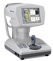
EM-3000  Product Detail

All-in-one Specular Microscope with “corneal endothelium photographing” and “automatic analysis”

Serial photographing of 15 shots

15 shots can be taken in series and errors during photographing are reduced. In addition, the best image among the 15 shots is automatically selected and displayed on the screen.

Wide photographing range and 7 capturing positions

Our unique technology enables a wide photographing range of 0.25 x 0.54 mm and allows you to observe the endothelium over a wide range. Images can be captured at 7 points: the center and 6 additional points (2, 4, 6, 8, 10, and 12-o’clock positions on a 6 mm arc). Because there are many photographing points, you can select the point with the best conditions even when the cornea surface is irregular. The cornea thickness is also measured at the same time.

Quick and automatic analysis of corneal endothelium cells

The software for automatic analysis is pre-installed, so images are analyzed automatically without using personal computers. Colorful icons and touch panel ensure easy operation for anyone.

Various display functions

The image of the corneal endothelium can be displayed with the cell shapes traced, as well as with different areas and structural forms of cells displayed in different colors. This provides a visual understanding of the condition of the corneal endothelium.

USB connector for printer and LAN connector for PC
  • USB-D connector: Connected to a Pict Bridge compatible printer to print images of the corneal endothelium and analysis results

  • USB-H connector: Connected to a barcode reader or electromagnetic card reader to enter patient ID data. A digital printer may also be connected.

  • LAN connector: After installing the “Data Transfer” software provided with the EM-3000 in your personal computer, inspection result files assigned a patient ID can be saved in the personal computer.

Specifications:
  • Observation and analysis of corneal endothelium (A-scan)

    • Photographing method: Non-contact

    • Photographing range: 0.54 (Y) x 0.25 (X) mm

    • Measurement mode: Auto / Manual 1 / Manual 2

    • Capturing position: Center +6 peripheral points

    • Cornea thickness measurement accuracy: +/- 10 mm

    • Analysis method: Automatic analysis

    • Analysis values

      • Number (the number of analyzed cells)

      • CD (cell density)AVG (average cell area)

      • SD (standard deviation of cell area)

      • CV (coefficient of variation of cell area)

      • Max (maximum cell area) Min (minimum cell area)

    • Histogram

      • Area (Polymegathism: Distribution by areas)

      • Apex (Plemorphism: Distribution by polygonal shapes types)

    • Main Unit

      • Display: 8.4" color LCD monitor

      • Stroke of moving parts: X-axis: 88 mm; Y-axis: 40 mm, Z-axis: 50 mm

      • Stroke of chin rest: 70 mm

      • Data output type: Printer / LAN / USB

      • Dimensions and weight: 308 (W) x 493 (D) x 453 (H) mm; Approx. 18 kg

      • Power source: 100 VAC-240 VAC; 50/60 Hz; 100 VA-130 VA


    Request Info

Enlarged Picture


Back


Join the Life Medical Equipment mailing list
Email:

 7874 NW 64th Street,  Miami, Florida, 33166
Tel: (305) 594-0000 - Fax: (305) 594-2020 -
© Copyright 2007 - Life Medical Equipment International Inc.